

Breathing Center Of The Brain
| Respiratory center | |
|---|---|
 Respiratory groups in the respiratory center and their influence | |
| Identifiers | |
| MeSH | D012125 |
| Anatomical terminology [edit on Wikidata] | |
The respiratory center is located in the medulla oblongata and pons, in the brainstem. The respiratory center is fabricated up of three major respiratory groups of neurons, two in the medulla and one in the pons. In the medulla they are the dorsal respiratory group, and the ventral respiratory grouping. In the pons, the pontine respiratory group includes two areas known as the pneumotaxic middle and the apneustic center.
The respiratory centre is responsible for generating and maintaining the rhythm of respiration, and also of adjusting this in homeostatic response to physiological changes. The respiratory center receives input from chemoreceptors, mechanoreceptors, the cerebral cortex, and the hypothalamus in lodge to regulate the rate and depth of animate. Input is stimulated by contradistinct levels of oxygen, carbon dioxide, and blood pH, by hormonal changes relating to stress and anxiety from the hypothalamus, and also past signals from the cerebral cortex to give a conscious command of respiration.
Injury to respiratory groups can cause various animate disorders that may require mechanical ventilation, and is normally associated with a poor prognosis.
Respiratory groups [edit]
The respiratory middle is divided into three major groups, two in the medulla and one in the pons. The ii groups in the medulla are the dorsal respiratory group and the ventral respiratory grouping. In the pons, the pontine respiratory group is fabricated up of ii areas – the pneumotaxic centre and the apneustic centre. The dorsal and ventral medullary groups control the basic rhythm of respiration.[one] [2] The groups are paired with ane on each side of the brainstem.[3]
Dorsal respiratory group [edit]
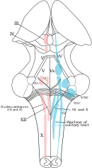
Solitary tract nucleus in the dorsal respiratory group and nucleus ambiguous of the ventral respiratory group shown in their positions on the medulla oblongata.
The dorsal respiratory grouping (DRG) has the about fundamental function in the control of respiration, initiating inspiration (inhalation). The DRG is a collection of neurons forming an elongated mass that extends most of the length of the dorsal medulla. They are most to the central canal of the spinal string, and just behind the ventral grouping. They set and maintain the rate of respiration.[four] [5]
Virtually of the neurons are located in the nucleus of the alone tract. Other of import neurons are constitute in the adjacent areas including the reticular substance of the medulla. The solitary nucleus is the end-betoken for sensory data arriving from the pontine respiratory grouping, and from ii cranial nerves – the vagus nerve, and the glossopharyngeal nerve. The lone nucleus sends signals to the respiratory centre from peripheral chemoreceptors, baroreceptors, and other types of receptors in the lungs in particular the stretch receptors. Thus, the dorsal respiratory group is seen every bit an integrating centre that gives the ventral respiratory group output to change the breathing rhythm.[4] [5]
Ventral respiratory grouping [edit]
In the medulla, the ventral respiratory grouping (VRG) consists of four groups of neurons that brand upward the exhalation (expiratory) area of respiratory control. This surface area is in the ventrolateral part of the medulla, about v mm inductive and lateral to the dorsal respiratory group. The neurons involved include those in the nucleus ambiguus, the nucleus retroambiguus, and the interneurons in the pre-Bötzinger circuitous.
The VRG contains both inspiratory and expiratory neurons.[half-dozen] [4] The ventral respiratory group of neurons are agile in forceful breathing and inactive during placidity, restful respirations.[1] The VRG sends inhibitory impulses to the apneustic center.
Pontine respiratory group [edit]
In the pontine tegmentum in the pons, the pontine respiratory group (PRG) includes the pneumotaxic and apneustic centers. These have connections between them, and from both to the lone nucleus.[7]
Pneumotaxic heart [edit]
The pneumotaxic heart is located in the upper part of the pons. Its nuclei are the subparabrachial nucleus and the medial parabrachial nucleus.[8] The pneumotaxic centre controls both the charge per unit and the pattern of breathing. The pneumotaxic center is considered an adversary to the apneustic centre, (which produces abnormal breathing during inhalation) cyclically inhibiting inhalation. The pneumotaxic middle is responsible for limiting inspiration, providing an inspiratory off-switch (IOS).[ix] It limits the burst of action potentials in the phrenic nerve, effectively decreasing the tidal volume and regulating the respiratory rate. Absenteeism of the center results in an increase in depth of respiration and a decrease in respiratory rate.
The pneumotaxic heart regulates the amount of air that can be taken into the body in each breath. The dorsal respiratory group has rhythmic bursts of action that are constant in duration and interval.[10] When a faster charge per unit of breathing is needed the pneumotaxic heart signals the dorsal respiratory group to speed upwardly. When longer breaths are needed the bursts of activity are elongated. All the information that the body uses to help respiration happens in the pneumotaxic centre. If this was damaged or in any fashion harmed information technology would brand breathing about impossible.
Ane study on this subject was on anaesthetized paralyzed cats earlier and after bilateral vagotomy. Ventilation was monitored in awake and anaesthetized cats breathing air or CO2. Ventilation was monitored both before and after lesions to the pneumatic centre region and after subsequent bilateral vagotomy. Cats with pontine lesions had a prolonged inhalation duration.[11] In cats, after amazement and vagotomy, pontine transaction has been described equally evoking a long sustained inspiratory discharges interrupted past brusk expiratory pauses.[ jargon ] In rats on the other hand, later anaesthesia, vagotomy and pontine transaction, this animate pattern was not observed, either in vivo or in vitro. These results propose interspecies differences between rat and cat in the pontine influences on the medullary respiratory middle.[12]
Apneustic centre [edit]
The apneustic centre of the lower pons appears to promote inhalation by constant stimulation of the neurons in the medulla oblongata. The apneustic heart sends signals to the dorsal group in the medulla to delay the 'switch off, the inspiratory off switch (IOS) indicate of the inspiratory ramp provided by the pneumotaxic centre. Information technology controls the intensity of breathing, giving positive impulses to the neurons involved with inhalation. The apneustic centre is inhibited past pulmonary stretch receptors and as well by the pneumotaxic centre. It likewise discharges an inhibitory impulse to the pneumotaxic centre.
Respiratory rhythm [edit]
Breathing is the repetitive procedure of bringing air into the lungs and taking waste products out. The oxygen brought in from the air is a abiding, on-going need of an organism to maintain life. This need is still there during sleep so that the functioning of this procedure has to exist automatic and be part of the autonomic nervous system. The in-breath is followed by the out-jiff, giving the respiratory bicycle of inhalation and exhalation. There are iii phases of the respiratory wheel: inspiration, post-inspiration or passive expiration, and late or active expiration.[xiii] [fourteen]
The number of cycles per infinitesimal is the respiratory rate. The respiratory charge per unit is set in the respiratory eye by the dorsal respiratory group, in the medulla, and these neurons are by and large concentrated in the solitary nucleus that extends the length of the medulla.[4]
The basic rhythm of respiration is that of tranquillity, restful breathing known equally eupnea. Serenity breathing but requires the activity of the dorsal group which activates the diaphragm, and the external intercostal muscles. Exhalation is passive and relies on the elastic recoil of the lungs. When the metabolic need for oxygen increases, inspiration becomes more forceful and the neurons in the ventral group are activated to bring about forceful exhalation.[1] Shortness of breath is termed dyspnea – the opposite of eupnea.
Clinical significance [edit]
Depression of the respiratory middle tin can be caused past: brain trauma, brain damage, a encephalon tumour, or ischemia. A depression can also exist caused past drugs including opioids, and sedatives.
The respiratory middle can be stimulated past amphetamine, to produce faster and deeper breaths.[15] Normally at therapeutic doses, this outcome is not noticeable, but may exist evident when respiration is already compromised.[15]
Encounter also [edit]
- Control of respiration
- Cough center
- Gag reflex
References [edit]
- ^ a b c Tortora, G; Derrickson, B (2011). Principles of beefcake & physiology (13th. ed.). Wiley. pp. 906–909. ISBN9780470646083.
- ^ Pocock, Gillian; Richards, Christopher D. (2006). Human physiology : the basis of medicine (3rd ed.). Oxford: Oxford University Press. p. 332. ISBN978-0-19-856878-0.
- ^ Saladin, Kenneth (2012). Anatomy Physiology The Unity of Grade and Function. pp. 868–871. ISBN9780073378251.
- ^ a b c d Hall, John (2011). Guyton and Hall textbook of medical physiology (twelfth ed.). Philadelphia, Pa.: Saunders/Elsevier. pp. 505–510. ISBN978-1-4160-4574-viii.
- ^ a b Saladin, Thousand (2011). Human anatomy (3rd ed.). McGraw-Hill. pp. 646–647. ISBN9780071222075.
- ^ Koeppen, Bruce M.; Stanton, Bruce A. (eighteen January 2017). Berne and Levy Physiology E-Book. Elsevier Health Sciences. ISBN9780323523400.
- ^ Vocal, Grand; Poon, CS (15 Nov 2004). "Functional and structural models of pontine modulation of mechanoreceptor and chemoreceptor reflexes". Respiratory Physiology & Neurobiology. 143 (2–3): 281–92. doi:10.1016/j.resp.2004.05.009. PMID 15519561. S2CID 38265906.
- ^ Song, Gang; Yu, Yunguo; Poon, Chi-Sang (2006). "Cytoarchitecture of Pneumotaxic Integration of Respiratory and Nonrespiratory Information in the Rat". Journal of Neuroscience. 26 (ane): 300–ten. doi:10.1523/JNEUROSCI.3029-05.2006. PMC6674322. PMID 16399700.
- ^ Dutschmann, Thou; Dick, TE (October 2012). "Pontine mechanisms of respiratory command". Comprehensive Physiology. ii (four): 2443–69. doi:10.1002/cphy.c100015. PMC4422496. PMID 23720253.
- ^ Dutschmann, Mathias (2011). Comprehensive Physiology. [Bethesda, Md.]: John Wiley and Sons. ISBN978-0-470-65071-4.
- ^ Gautier, H; Bertrand, F (1975). "Respiratory furnishings of pneumatic heart lesions and subsequent vagotomy in chronic cats". Respiration Physiology. 23 (i): 71–85. doi:10.1016/0034-5687(75)90073-0. PMID 1129551.
- ^ Monteau, R.; Errchidi, S.; Gauthier, P.; Hilaire, M.; Rega, P. (1989). "Pneumotaxic centre and apneustic animate: Interspecies differences between rat and true cat". Neuroscience Letters. 99 (3): 311–6. doi:ten.1016/0304-3940(89)90465-5. PMID 2725956. S2CID 42790256.
- ^ Mörschel, Thou; Dutschmann, K (12 September 2009). "Pontine respiratory activity involved in inspiratory/expiratory phase transition". Philosophical Transactions of the Royal Society of London. Serial B, Biological Sciences. 364 (1529): 2517–26. doi:ten.1098/rstb.2009.0074. PMC2865127. PMID 19651653.
- ^ Ramirez, JM; Dashevskiy, T; Marlin, IA; Baertsch, N (December 2016). "Microcircuits in respiratory rhythm generation: commonalities with other rhythm generating networks and evolutionary perspectives". Electric current Opinion in Neurobiology. 41: 53–61. doi:10.1016/j.conb.2016.08.003. PMC5495096. PMID 27589601.
- ^ a b Westfall DP, Westfall TC (2010). "Miscellaneous Sympathomimetic Agonists". In Brunton LL, Chabner BA, Knollmann BC (eds.). Goodman & Gilman'southward Pharmacological Basis of Therapeutics (twelfth ed.). New York, The states: McGraw-Hill. ISBN9780071624428.
Further reading [edit]
- Levitzky, Michael G. (2002). Pulmonary Physiology (sixth ed.). McGraw-Hill Professional. pp. 193–iv. ISBN978-0-07-138765-1.
- Costanzo, Linda S. (2006). Physiology (3rd ed.). Philadelphia, PA: Elsevier. p. 224. ISBN978-one-4160-2320-iii.
- Shannon, Roger; Baekey, David Thousand.; Morris, Kendall F.; Nuding, Sarah C.; Segers, Lauren Southward.; Lindsey, Bruce G. (2004). "Pontine respiratory grouping neuron discharge is altered during fictive cough in the decerebrate true cat". Respiratory Physiology & Neurobiology. 142 (1): 43–54. doi:10.1016/j.resp.2004.05.002. PMID 15351303. S2CID 8425115.

Breathing Center Of The Brain,
Source: https://en.wikipedia.org/wiki/Respiratory_center
Posted by: handyowly1985.blogspot.com
0 Response to "Breathing Center Of The Brain"
Post a Comment